The ordeal for those to go under the (laser) knife begins with thorough evaluations to pinpoint any major design flaws, or life-hazards incurred in their basic cerebral mechanics. For the thirteen epilepsy patients involved in initial trials based at the Emory University School of Medicine, this included any number of unique labors and privileges. High on the list were the use of subdural (under the scalp, on the brain surface) electrode arrays to localize where seizures first begin. At its most basic level this would mean which half of the brain is primarily to blame. For those outfitted with this gear, cortical areas involved in vital functions like speech or face recognition can be also predetermined. These regions, whimsically designated as the “eloquent cortex” by neurosurgeons, are not so much the parts of the cortex that might be merely missed if ablated, but rather those whose loss could not be ignored.
Several patients were additionally given what is known as a “Wada” test to expand and confirm the electrode evaluations. This diabolical thrill involves injecting either the left or right arterial blood feedstock with a powerful barbiturate to see what critical functions drop out, or temporarily go offline. The whole patient workup is supplemented by MRI and PET scans to get a feel for the anatomy at hand; more-or-less a dry run of the procedure about to go down. This typically involves further dubious ingestion of radioactive labels in the case of PET, and gadolinium contrast agents for the MRI. In this vein of patient precautions, we should also make mention of the smattering of X-rays you may later absorb during the procedure from the fluoroscopic arm used to check on the probe position.
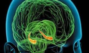
The MRI is also critical for the placement of the laser probe, which is inserted along a minimally-invasive trajectory, beginning from the back of the head. The angle of attack is chosen such that the probe inserts in line with the long axis of the hippocampus, the region of the brain that along with the adjacent amygdala is the primary surgical target (see the orange regions in the image above). This part is a bit tricky because the hippocampus is close to several critical components in the vision chain. One patient in the trial incurred a significant loss in a discrete patch of their visual field when an anchor screw that secures and aligns the apparatus was improperly threaded.
A preoperative diagram showing the proposed laser trajectory.
The optical fiber is capped with a diffuser that disperses the 980-nm laser beam. At this wavelength (infrared) cells essentially experience the light as heat which destroys them. In the actual procedure the tip of the fiber is slowly backed out of its bore, unloading its energy like a commando spraying bullets as he retreats from an enemy cave. The whole point of the laser concept is to avoid the gross tissue damage you would otherwise get with brute force approaches. Typically this would entail either scooping out brain in small ice cream-like dollops, or melting it with ultrasonics or perhaps an indiscriminate RF-emitting antenna. In order to avoid inflicting the same kind of trauma with an errant heat wand, the surgeons have come up with a solution extraordinaire.
A remarkable refinement of the standard MRI lets the temperature of discrete parts of the brain be remotely interrogated. Known as MRI thermometry (MRIT), the technique capitalizes on the temperature sensitivity of the proton resonance frequency of water molecules. On a more technical end, the thermal coefficient of this resonance is independent of most tissue types and is linear over the ranges of interest. (The actual temperature sensitivity that can be achieved is about 0.2 degrees C). On the lower tech end, the technique was optimized using a commercially available tofu tissue phantom supplied by a local supermarket. Tofu has a density, speed of sound, and attenuation coefficient similar to brain tissue making it the go to material for MRTI development. MRTI initially proved itself as a way to quantify ultrasonic heating for advanced applications like localized drug release deep within tissue.
www.extremetech.com